


A 3D skin analysis machine detects a comprehensive range of issues: wrinkles, pores, pigmentation, acne, hydration, oil levels, UV damage, and elasticity. It uses AI to evaluate multiple key parameters, moving far beyond surface-level observation. Clinics and spas trust this technology for precise assessment and treatment tracking. Research shows digital analyzers achieve 92–96% accuracy for pigmentation and 91% for pore size, compared to 70% and 55% with visual inspection. The system also offers 95% test-retest reliability. In the following sections, I break down each detection category in detail.
Key Takeaways
●A 3D skin analysis machine reveals hidden sun damage and UV spots long before they become visible on your skin.
●It measures exact numbers for wrinkle depth, pore size, and pigmentation density, replacing guesswork with objective data.
●You can track treatment progress with precise readings on hydration, oil levels, and elasticity across multiple visits.
●The machine identifies acne triggers by measuring bacterial activity and inflammation levels with high accuracy.
●Precise 3D imaging measures scar volume and depth, helping you monitor the effectiveness of treatments over time.
Texture and Topography in 3D Skin Analysis
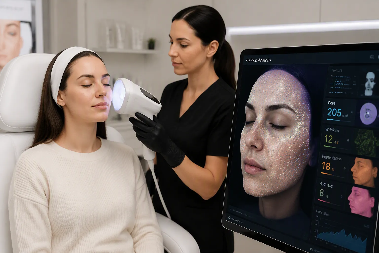
I rely on the 3D skin analysis machine to capture the exact depth and contour of every feature on your face. This technology reveals details invisible to the naked eye. Two areas benefit most from this precision: wrinkles and pores.
Wrinkles and Fine Lines
I measure the maximum depth of your glabellar region while you frown at a distance of about 250 mm. The laser-based system demonstrates excellent geometric measurement accuracy. It achieves correlation coefficients of r = 0.9998 for surface area, 0.9995 for perimeter, 0.9995 for volume, and 0.9928 for depth. Measurement errors stay within 1–2% for area, perimeter, and volume, and within ±0.5 mm for depth. This reliability allows me to detect fine topographic variations.
Different technologies make these measurements possible. The Antera 3D reconstructs skin using shape-from-shading and photometric stereo measurements across seven visible wavelengths. The DermaTOP system uses interference fringe projection profilometry to collect 3D information on skin topography. The SWIRL method quantifies five wrinkle parameters: number, length, width, area, and depth. The Wrinkle Gray Scale numerically measures the brightness difference between wrinkles and surrounding skin — more pronounced wrinkles produce higher gray values.
I see 3D-measured glabellar depth show moderate positive correlations with clinician-rated severity (r = 0.5327) and participant-reported severity (r = 0.5855). One representative case stands out: measurable improvement in 3D-derived wrinkle depth appeared even when clinical severity grading showed minimal change. This highlights the increased sensitivity of quantitative 3D skin analysis over ordinal visual assessment.
Pores and Scarring Depth
A cross-sectional study used the Primos-CR® 3D imaging system to measure skin surface profiles from the cheeks of 101 Korean women aged 3–68 years. This system offers a resolution of 20 μm to plane and 5 μm to depth, enabling precise measurement of skin pores. The study classified pores into five groups based on size, density, and elongation using K-means clustering. Results showed pores become larger over time, with adjacent pores connecting in elderly subjects. 3D imaging overcomes 2D analysis limitations by providing topographical information inside pores.
The machine can also measure the volume and depth of scars. This quantifiable data helps track treatment progress over time.
Pigmentation and Color Detection
Color variation across your skin tells a detailed story. I use spectral analysis to read that story with precision. The system examines your skin under multiple light wavelengths. This approach reveals pigment patterns that sit deep below the surface.
Sunspots, Freckles, and Melasma
I can distinguish a wide range of pigmentation disorders with this technology. The table below shows what the system identifies.
| Pigmentation Disorder | How the Machine Detects It |
|---|---|
| Melasma | UV damage diagnosis and spectral analysis |
| Chloasma | Facial skin analyzer detection |
| Freckles | Sun spot identification under intense UV exposure |
| Vitiligo | Eight spectral analysis techniques |
| Age spots | UV damage diagnosis |
| Sun spots | Epidermal melanin level analysis |
| Birthmarks | Direct detection |
| Hemangiomas | Direct detection |
| Port-wine stains | Direct detection |
The AI then quantifies pigmentation density. I compare your readings against baseline data. This gives me an objective measure of pigment concentration. I track how these levels shift across visits. A fading sunspot shows a clear drop in melanin density. Melasma patches often respond differently. The AI captures those subtle changes too.
Redness and Vascular Lesions
Redness intensity is another core measurement. I map vascular activity across your face. The system highlights areas of increased blood flow. Rosacea appears as concentrated zones of elevated redness. Hemangiomas and port-wine stains show distinct vascular signatures. I measure the intensity of each lesion. This helps me separate active inflammation from stable vascular features.
The AI assigns numerical values to redness. I use these values to build a treatment plan. A high redness score near your cheeks may point to rosacea triggers. A stable score suggests a benign feature. Over time, I compare scores to see if your skin is calming down or flaring up. This objective tracking removes guesswork from your skincare routine. A 3D skin analysis gives you clear numbers instead of vague impressions.
Blemishes and Acne Analysis
Blackheads and Whiteheads
I use the 3D skin analysis system to identify every type of blemish on your skin. The machine separates surface spots from deeper concerns. This distinction matters for your treatment plan.
| Blemish Type | What It Means |
|---|---|
| Spots | Surface pigmentation like sun spots, freckles, acne marks, and moles, seen under standard white light |
| UV Spots | Hidden pigment beneath the epidermis, revealed with 365nm ultraviolet light |
| Brown Spots | Deeper spots such as chloasma and freckle-like nevi that may rise to the surface over time |
| Red Zone | Capillary conditions like inflammatory acne, rosacea, and spider nevi |
| Porphyrin | Bacterial metabolites on hair follicles that fluoresce under special lighting |
Blackheads and whiteheads sit at the surface. I detect them through standard white light imaging. The system maps each pore opening and measures blockage density. This gives me a clear count of active comedones.
Inflammation and Bacterial Activity
Porphyrin levels tell me how active bacteria are on your skin. Porphyrins are bacterial activity markers. They indicate acne-prone conditions. The machine measures the porphyrin ratio present in your skin. This reading shows me where inflammation may flare up next.
I also check fluorescent agent detection. This step uncovers hidden residues on your skin. The red zone reading captures capillary conditions. Inflammatory acne, rosacea, and spider nevi all appear here. I detect them through hemoglobin in the dermal papillary layer. A high red zone score points to active inflammation. A stable score suggests your skin is calm.
These measurements remove guesswork from acne treatment. I track your porphyrin ratio across visits. A falling ratio means the bacteria are under control. A rising ratio warns me to adjust your routine. You get clear numbers instead of vague impressions about your skin.
Hydration, Oil, and Elasticity
Moisture and Sebum Levels
I measure how much water sits in your outer skin layer and how much oil your glands produce. The machine reads both values at the same time. A low moisture score tells me your barrier may be compromised. A high sebum reading points to overactive oil production. I compare these two numbers to classify your skin type with real data instead of a guess.
The system tracks these readings across visits. If your moisture rises after a new serum, I see the change in numbers. If your oil output drops after a treatment, the machine confirms it. This feedback loop keeps your routine honest. You stop wondering whether a product works. You watch the trend line move.
Skin Firmness and Sagging
I assess firmness and elasticity through displacement mapping. The system reconstructs real elastin fiber networks from your dermis using 3D confocal imaging. It embeds the segmented fiber geometry into a finite element mesh of the surrounding dermal matrix. Each fiber becomes a 3D beam element that captures its individual shape and orientation. A quasi-static unconfined compression simulation then runs with boundary conditions constraining the horizontal, vertical, and depth directions. The model compresses to 80% of its original height, a 20% compression. I read the stress-strain response to compute two values. Firmness is your skin’s resistance to deformation under load. Elasticity is its ability to return to its original shape after that load releases. Elastin drives the initial low stiffness you feel before stiffer collagen fibers engage, and it stretches far more than collagen does.
Sagging follows a different pathway. Collagen degradation contributes to sagging, and 3D imaging can assess the collagen-related features behind it. Senescent fibroblasts increase ECM degradation through MMP activation. That process reduces collagen density, thickness, and organization. Collagen breakdown, driven by rising MMPs and falling TIMPs, accelerates skin aging. These ECM changes link to reduced elasticity, more wrinkles, and visible sagging. Clinical evaluations confirm significant age-related reductions in skin elasticity, firmness, and density, along with increased wrinkles and decreased plumpness. Advanced methods like 3D imaging let me investigate these microstructures in detail. A 3D skin analysis gives me the objective baseline I need to track whether your firmness holds steady or declines over time.
UV Damage and Hidden Concerns
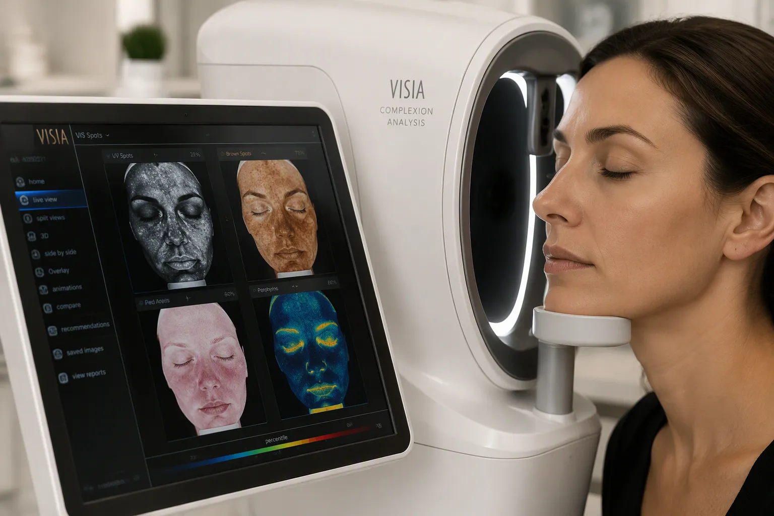
Sun Damage and UV Spots
I use this machine to reveal sun damage that stays invisible to your naked eye. The system uses 365nm ultraviolet light to reveal hidden pigment spots beneath the epidermis. This exposes melanin distribution and subsurface damage. I can detect invisible sun damage well before it reaches the epidermis.
| Hidden UV Damage Type | Description |
|---|---|
| UV Spots | Areas just below the skin surface damaged by sun exposure |
| Brown Spots | Pigmentation and discoloration below the surface that may become visible later |
| Bacterial Presence (Porphyrins) | Bacteria within pores exposed by UV lighting, helping predict future skin issues |
I track these hidden spots across visits. A fading UV spot confirms your sun protection routine works. A growing spot warns me to adjust your approach before visible damage forms. This preventive strategy saves you time and money. You address problems before they surface.
Sensitivity and Vascular Health
I also assess skin sensitivity through vascular health markers. The system can detect capillary conditions including hemangiomas, port-wine stains, and other vascular lesions. It identifies areas of increased blood flow and inflammation, helping differentiate between active rosacea and stable vascular features. The red zone reading captures capillary conditions like inflammatory acne, rosacea, and spider nevi. These measurements help me build a complete picture of your skin health. I combine sensitivity scores with UV damage readings. This gives me a full map of hidden threats. Your 3D skin analysis delivers actionable data instead of guesswork.
I have walked you through five detection categories: texture and topography, pigmentation and color, blemishes and acne, hydration and elasticity, and UV damage with vascular health. Each category gives you hard numbers. You see wrinkle depth in millimeters. You see melanin density scores. You see porphyrin ratios and redness values. These measurements build a complete picture of your skin’s unique profile.
This objective data changes how you track treatment results. A serum either moves your moisture score or it does not. A laser either reduces your scar volume or it does not. You stop guessing. You watch the numbers.
I encourage you to book a professional 3D skin analysis for your next skincare consultation. Personalized insights lead to better decisions. Your skin deserves that clarity.
FAQ
How accurate is a 3D skin analysis machine compared to visual inspection?
Digital analyzers achieve 92–96% accuracy for pigmentation and 91% for pore size. Visual inspection reaches only 70% and 55% for those same measures. The system also offers 95% test-retest reliability. These numbers show why clinics trust this technology over the naked eye.
Can the machine detect skin issues that are not yet visible?
Yes. The system uses 365nm ultraviolet light to reveal hidden UV spots beneath the epidermis, as well as identify clogged pores and other early signs of skin damage.
What skin conditions can the machine identify beyond wrinkles and pores?
The system detects melasma, chloasma, freckles, vitiligo, birthmarks, hemangiomas, port-wine stains, and rosacea. It also measures porphyrin levels to show bacterial activity. Red zone readings capture inflammatory acne and spider nevi. Each condition gets a numerical score for objective tracking.
How does the machine measure skin elasticity and firmness?
The system reconstructs real elastin fiber networks from your dermis using 3D confocal imaging. It runs a compression simulation to read your stress-strain response. Firmness is your skin’s resistance to deformation. Elasticity is its ability to return to shape after that load releases.
What makes this technology better than a traditional skin consultation?
A traditional consultation relies on visual assessment and subjective judgment. This machine gives you hard numbers for wrinkle depth, melanin density, and moisture levels. You track treatment results across visits. A serum either moves your score or it does not. You stop guessing.
Post time: Sep-23-2026


