


I use 3D skin analysis to examine skin conditions with exceptional detail. This technology relies on stereoscopic imaging and advanced algorithms. It builds a high-resolution 3D model of your skin. The system measures depth, volume, and surface irregularities. This non-invasive method offers a precise roadmap. I can identify specific features like spots, wrinkles, texture, pores, brown spots, and red areas. UV filters map pigment below the surface. This reveals hidden sun damage before it becomes visible. Understanding these steps helps you appreciate the technology.
Key Takeaways
●3D skin analysis uses multiple cameras and UV light to create a detailed 3D model of your skin.
●It detects spots, wrinkles, pores, and sun damage hidden beneath the surface.
●The system measures skin features with high precision and tracks changes over time.
●Doctors use this technology to plan personalized treatments and monitor progress.
●You can see your skin health objectively and take charge of your skincare routine.
How 3D Skin Analysis Captures High-Resolution Images
Stereoscopic Imaging and Multi-Angle Capture
I start the process by positioning multiple cameras and sensors around your face. Each camera captures the skin from a different angle. This multi-angle approach provides the raw data needed for 3D reconstruction. The system combines ultraviolet (UV) and visible light through multiple LED arrays to create a three-dimensional effect. UV filters play a critical role here. They map pigment concentration below the surface and show where sun damage is forming.
UV light is absorbed by epidermal melanin located just beneath the skin surface. This absorption enables the visualization of UV spots caused by sun damage. These spots would otherwise remain hidden under visible light alone. The difference between visible light and UV-filtered analysis is substantial.
| Aspect | Visible Light Alone | UV-Filtered 3D Analysis |
|---|---|---|
| Detection depth | Surface only | Surface and subsurface |
| Sun damage visibility | A few visible dark spots | Subsurface UV damage detectable years before it surfaces |
| Concealed conditions | Not detectable by mirror or magnifying lamp | Identifies concealed skin conditions invisible to the unaided eye |
UV light exposes sun damage and pigmentation beneath the surface that is not yet visible.
This capability makes UV imaging particularly useful for diagnosing and treating melasma. It accurately identifies active and potential problematic pigmentation, including latent pigmentation such as unoxidised melanin.
Lighting and Calibration Techniques
The system employs various lighting modes to capture both surface and subsurface details. These modes include IntelliFlash (standard white light), cross-polarized light, and ultraviolet (UV) light. Each mode serves a specific purpose in the capture process.
Cross-polarized light reduces glare and highlights subsurface features. It penetrates just below the skin surface to assess attributes such as redness and rosacea. This glare reduction enables evaluation of subsurface skin irregularities. Parallel polarized light strengthens surface optical reflection. It suppresses subsurface scattering that can wash out surface detail. This enhances clarity of surface texture and allows clearer observation of skin flatness, fine lines, and wrinkles. The skin lines become easier to distinguish, which supports AI-based wrinkle counting.
Proper calibration ensures repeatable measurements across sessions. I rely on these lighting techniques to build a detailed roadmap of your skin condition.
How Software Reconstructs a 3D Skin Model
Generating a 3D Mesh from Image Data
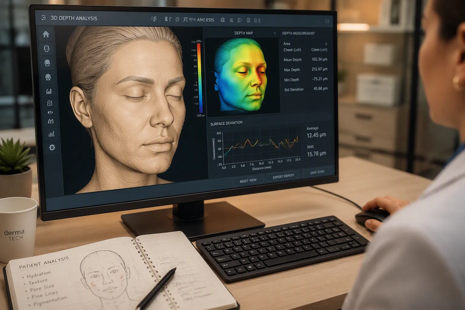
After capturing images from multiple angles, the software begins its work. It uses algorithms to stitch the raw data together. This process creates a high-resolution three-dimensional model of the skin. The model becomes the foundation for personalized dermatological treatment.
Hair and eyebrow regions create a challenge. Without special handling, these areas produce artifacts in the mesh model. I rely on several techniques to solve this problem. Depth-gating prevents Gaussians from representing the underlying skin during optimization. This ensures only facial hair strands appear in the capture. Grounding strands snap reconstructed hair to the tracked face mesh at their root positions. This eliminates floating or disconnected geometry.
For eyebrows specifically, the system resolves root-tip ambiguity. It uses a side-aware rule and an angular threshold of 30 degrees. This correctly assigns which endpoint is the root. It avoids inverted or misaligned eyebrow strands on the mesh.
Sparsity-aware refinement also plays a role. Redundant or nearly-parallel strands get pruned and redistributed under multi-view supervision. This prevents over-dense reconstructions from bleeding into the skin.
The method separates skin from non-skin regions on 3D head scans. It fuses multi-view image features with 3D geometric features. This segmentation prevents the template mesh from being pulled toward noisy scan surfaces in hair regions. The approach improves registration accuracy by 8.89% over purely 2D methods and 14.3% over purely 3D methods.
Calculating Depth and Surface Irregularities
Once the software builds a clean mesh, it calculates depth and surface irregularities. The system compares the 3D skin model to a smooth reference surface. Every deviation becomes a measurable feature.
Wrinkles appear as linear depressions in the mesh. The software measures their depth. Pores show as small circular indentations. The system calculates their diameter. Texture roughness emerges as overall surface variation across the skin.
The algorithms quantify these features with high precision. For example, the system can distinguish between fine lines and deep wrinkles based on their depth profile. This level of detail supports accurate diagnosis.
The 3D skin analysis system provides repeatable measurements across sessions. Clinicians can track changes over time with confidence. The software outputs a comprehensive skin report. It lists spots, wrinkles, texture, pores, brown spots, and red areas. This data directly supports personalized dermatological treatment. The skin model reveals conditions invisible to the naked eye. I use this information to guide treatment decisions.
How 3D Skin Analysis Measures Wrinkles, Pores, and Pigmentation
Assessing Wrinkle Depth and Pore Diameter
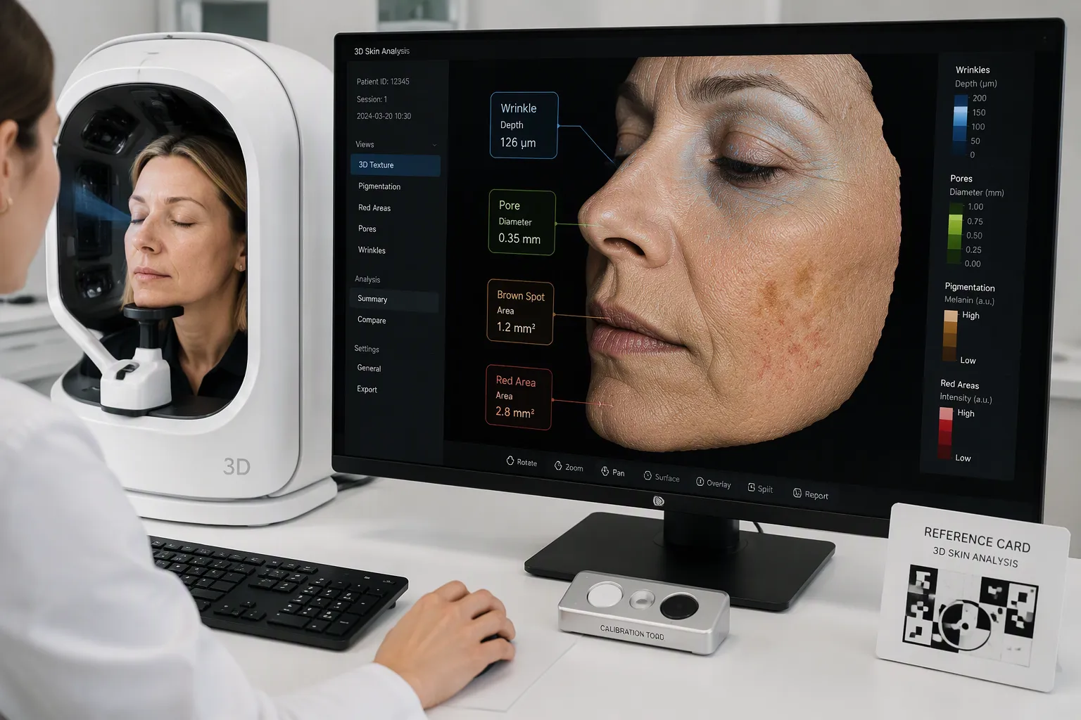
I measure wrinkle depth and pore diameter with high precision using the reconstructed 3D mesh. The software treats every depression in the skin surface as a measurable feature. Wrinkles appear as linear valleys in the mesh. The system calculates their depth by comparing each point to a smooth reference surface. Fine lines show shallow depth profiles. Deep wrinkles display pronounced depressions that extend further into the dermis.
Pore diameter follows a similar logic. Pores appear as small circular indentations scattered across the skin surface. The software detects each pore and measures its diameter. This measurement reveals pore size distribution across different facial zones. The nose and inner cheeks typically show larger pores than the outer cheeks and forehead.
The system identifies specific features with remarkable consistency. These features include spots, wrinkles, texture, pores, brown spots, and red areas. Each feature receives a quantitative score. I can compare these scores across treatment sessions to track improvement or deterioration.
Repeatability matters for clinical decision-making. The same lighting conditions and calibration settings ensure consistent measurements over time. A patient who returns after six weeks of treatment receives comparable data. This allows me to assess whether a topical retinoid reduced wrinkle depth or whether a pore-refining serum achieved its goal.
Clinical studies confirm strong agreement between 3D measurements and dermatologist ratings. This demonstrates the reliability of 3D skin analysis for telehealth applications. A dermatologist reviewing the 3D model and feature scores can make informed decisions without an in-person visit. The technology bridges geographic gaps in dermatological care.
Mapping Pigmentation and Texture Changes
Pigmentation mapping requires the UV-filtered imaging I described earlier. The system detects melanin concentration at different depths beneath the skin surface. Brown spots appear as areas of elevated melanin in the epidermal layer. Red areas indicate vascular activity or inflammation near the surface.
The software generates a pigmentation map that color-codes different concentrations. Darker regions represent higher melanin density. This map reveals sun damage that remains invisible under normal lighting. A patient might see a clear complexion in the mirror. The UV map shows scattered pigment deposits forming years before they surface.
Texture changes follow a different measurement path. The system calculates surface roughness by analyzing variations in the mesh geometry. Smooth skin shows minimal deviation from the reference surface. Rough texture displays frequent peaks and valleys across the measured area.
I combine pigmentation data with texture scores to build a complete skin profile. This profile guides treatment selection. A patient with significant UV damage and rough texture might benefit from a different protocol than someone with primarily vascular concerns.
The 3D skin analysis system outputs a comprehensive report after each session. This report lists every detected feature with its measurement. I use this data to design personalized dermatological treatment plans. The objective measurements remove guesswork from skincare recommendations.
Tracking changes over time reveals treatment efficacy. A series of sessions shows whether pigmentation faded or texture improved. Patients appreciate seeing their progress quantified. This visual and numerical feedback encourages adherence to skincare regimens.
Real-World Applications of 3D Skin Analysis
Clinical Diagnosis and Treatment Planning
I use 3D skin analysis in clinical settings to support diagnosis and treatment planning. The technology is non-invasive and fits naturally into scientific skin management workflows. Dermatologists can capture a detailed skin model before treatment begins. This baseline helps them plan procedures with greater confidence.
The evidence shows strong clinical value. Observers reported that 3D images improved their capacity to use the IGAIS scale. Higher ICC values confirmed greater perception of depth and volume. This led to more coherent judgments across raters. The percentage of improvement scored by raters matched 3D SPM findings. Inter-rater agreement was higher when ratings were based on 3D images.
| Rated Aspect | Dermatologists’ Rating | Supporting Detail |
|---|---|---|
| Quantifying post-treatment volumetric changes | Positive | 3D images improved IGAIS use; higher ICC confirmed better depth perception |
| Agreement between rater scores and 3D SPM | High | Improvement scores matched 3D SPM findings; inter-rater agreement was higher |
| Overall concordance and accuracy | Confirmed | Direct correlation between IGAIS and 3D SPM confirmed |
One limitation exists. Observers noted the IGAIS scale restricted evaluation to zero or positive values. Negative outcomes were detected only by 3D SPM. This reduced correlation for adverse outcomes. Despite this, raters overestimated improvement in a nontreatment control. The technology still proved precise in detecting absence of intervention.
Personalized Skincare Regimens
I build personalized skincare regimens using the detailed data from each session. The system identifies spots, wrinkles, texture, pores, brown spots, and red areas. Each feature receives a quantitative score. I compare these scores across sessions to track progress.
Patients see their skin health measured objectively. This removes guesswork from product selection. A patient with UV damage and rough texture receives a different protocol than someone with vascular concerns. The data guides every recommendation.
Clinics use these machines for scientific skin management. Cosmetic brands use them to support product recommendations. The technology serves both medical and retail settings.
I find that patients adhere better to regimens when they see quantified results. A six-week follow-up shows whether a retinoid reduced wrinkle depth. It reveals whether a serum improved texture. This feedback loop keeps patients engaged. The non-invasive nature makes regular monitoring practical. No downtime or discomfort interferes with daily life.
I trust 3D skin analysis because clinical standards validate its measurements. The system produces repeatable results across sessions. This reliability makes it a dependable tool for tracking skin health over time.
The future looks promising. AI integration will sharpen diagnostic accuracy. Real-time tracking will let you monitor changes daily. Home devices will bring this technology into your bathroom.
I encourage you to view this tool as proactive skincare. It complements professional judgment. It does not replace your dermatologist. Use it to catch problems early. Use it to measure your progress. The technology empowers you to take charge of your skin health with confidence.
FAQ
Does 3D skin analysis hurt?
No. The process is non-invasive and captures images safely. Multiple cameras and sensors take pictures from different angles. I see no discomfort or downtime. You simply sit still while the system works.
What skin features does the system detect?
I measure spots, wrinkles, texture, pores, brown spots, and red areas. UV filters also map pigment below the surface. This reveals sun damage forming years before it becomes visible.
How accurate is 3D skin analysis?
Clinical studies confirm strong agreement between 3D measurements and dermatologist ratings. The system delivers repeatable results across sessions.
Can I track my skin changes over time?
Yes. The same lighting and calibration settings ensure consistent measurements every session. I compare wrinkle depth, pore diameter, and pigmentation scores across visits. This helps me assess whether treatment is working.
Post time: Sep-23-2026


