


Quick Answer
Professional face analysis machines rely on a layered stack of imaging technologies working in concert. Multi-spectral imaging captures skin at different wavelengths—visible, cross-polarized, parallel-polarized, and ultraviolet—to reveal pigment, vascularity, and surface texture. 3D depth sensing (structured light or stereo vision) reconstructs facial geometry for volume and symmetry assessment. AI-driven analysis then processes these data streams into quantified scores and visual reports. The global AI skin analysis market, valued at USD 1.02 billion in 2026, is projected to reach USD 2.45 billion by 2031 at a 19.3% CAGR, driven by clinical demand for objective, reproducible skin diagnostics. Understanding what each imaging layer actually does—and what it cannot do—is essential for buyers evaluating equipment claims.
Key Takeaways
- ✔ Multi-spectral imaging is the diagnostic core of professional skin analysis, with UV and cross-polarized light revealing subsurface conditions invisible under standard illumination.
- ✔ True 3D depth sensing requires dedicated hardware (structured light, stereo cameras, or time-of-flight sensors); “3D-like” software interfaces on 2D systems are not equivalent.
- ✔ Standardized capture conditions—fixed lighting, consistent head positioning, calibrated color references—determine whether longitudinal comparisons are clinically meaningful.
- ✔ ISO 13485 certification indicates a manufacturer’s quality management system meets medical device standards, a relevant benchmark for clinical-grade equipment.
- ✔ The MEICET Pro-D exemplifies multi-spectral 2D analysis with 50+ skin dimensions, while the MEICET S7 adds true 3D depth mapping for contour and volume assessment.
The Imaging Stack: Why One Sensor Cannot Do Everything
No single imaging technology captures the full picture of facial skin and structure. Professional face analysis machines therefore combine multiple acquisition modalities, each optimized for a specific diagnostic target. A standard professional session typically involves four to nine image captures per visit, each using a different illumination or polarization configuration.
The reason is physical. Skin is a turbid medium: light penetrates to varying depths depending on wavelength, and different tissue components absorb or scatter light differently. Melanin absorbs broadly across the visible spectrum, hemoglobin absorbs strongly in the green band, and collagen fluoresces under UV. Pores and fine lines alter surface reflectance geometry. Capturing all of these simultaneously with a single white-light photograph is impossible. The imaging stack solves this by interrogating the skin under controlled, sequential conditions.
The MEICET Pro-D system illustrates this approach in practice, offering 50+ skin analysis dimensions across blemishes, sensitivity, pigmentation, texture, and visible aging. The device’s clinical value derives not from any single sensor but from the coordinated analysis of multiple spectral views.
Multi-Spectral Imaging: The Diagnostic Backbone
Multi-spectral imaging is the most important technology for skin health assessment, enabling visualization of structures at different depths within the skin. Each spectral band targets specific chromophores and tissue properties. The table below summarizes the primary modes used in professional systems.
| Imaging Mode | Wavelength/Access | Primary Diagnostic Target | What It Reveals |
|---|---|---|---|
| Standard RGB | Visible white light | Surface appearance | Color, texture, visible lesions, overall complexion |
| Cross-Polarized (CPL) | Visible, crossed polarizers | Subsurface vascularity | Erythema, rosacea, telangiectasia, inflammation depth |
| Parallel-Polarized (PPL) | Visible, parallel polarizers | Surface texture | Pores, fine lines, wrinkles, skin roughness |
| UV / Wood’s Light | ~365 nm ultraviolet | Pigment and porphyrins | Sun damage, melasma depth, C. acnes fluorescence |
| Blue Light | ~405 nm | Sebum and acne | Porphyrin detection, oiliness distribution |
Cross-polarized imaging works by blocking surface-reflected light while transmitting light that has penetrated deeper into the dermis and returned after scattering. This reveals vascular structures and pigmentary changes beneath the stratum corneum. UV imaging excites porphyrins produced by Cutibacterium acnes bacteria, causing them to fluoresce orange-red—a direct visual indicator of acne activity that is invisible under normal light. Parallel-polarized imaging retains surface reflectance, making it the optimal mode for quantifying texture parameters like pore size and wrinkle depth.
The diagnostic value of multi-spectral imaging is well established in dermatological research. A 2026 review in Advanced Photonics Research documented that hyperspectral and diffuse optical imaging techniques, when paired with machine learning, have achieved sensitivities and specificities exceeding 90% for melanoma, basal cell carcinoma, and squamous cell carcinoma detection. While professional face analysis machines operate at a different clinical scope than diagnostic dermatology devices, the underlying principle—that spectral information beyond RGB improves diagnostic accuracy—is identical.
3D Depth Sensing: What It Adds and What It Requires
True 3D face analysis captures geometric data that 2D imaging cannot approximate, but only when the system uses dedicated depth-sensing hardware. The distinction matters because marketing language often conflates “3D visualization” (a software rendering of a 2D image) with “3D acquisition” (actual depth measurement).
Three hardware approaches dominate professional 3D face analysis:
- Structured light: Projects a pattern of dots or lines. A 2025 study in the Journal of Indian Prosthodontic Society reported mean discrepancies as low as 0.35 ± 0.33 mm.
- Stereo vision: Uses two or more cameras to triangulate depth, similar to human binocular vision.
- Time-of-flight (ToF): Measures the round-trip time of light pulses.
The MEICET S7 3D system employs structured-light acquisition combined with a 24MP ultra-micro lens and nine imaging modes, extending its utility from pure geometry into skin-quality assessment through 3D local texture analysis.
What 3D adds to the diagnostic picture: volumetric change in cubic millimeters, angular measurements of facial projection, and true three-plane symmetry assessment. For practices discussing dermal fillers, thread lifts, or facial contouring, these data points transform a subjective conversation into a quantifiable one.
Standardization and Repeatability: The Overlooked Technology
The most sophisticated imaging sensor produces unreliable data if capture conditions vary between visits. Standardization is not a sensor technology, but it functions as a critical enabling layer for longitudinal analysis.
Professional systems address standardization through several mechanisms. Fixed lighting geometry ensures that illumination intensity and angle remain constant across sessions. Mechanical head positioning—chin rests, forehead supports, or both—constrains the subject’s position to a reproducible coordinate frame. The MEICET Pro-D includes forehead and chin supports specifically for this purpose. Color calibration references correct for drift in sensor response or ambient light leakage.
AI Analysis: From Photons to Clinical Insight
AI algorithms transform raw spectral and geometric data into the quantified scores, visual overlays, and trend reports that clinicians actually use. The imaging hardware captures photons; the AI layer determines what those photons mean.
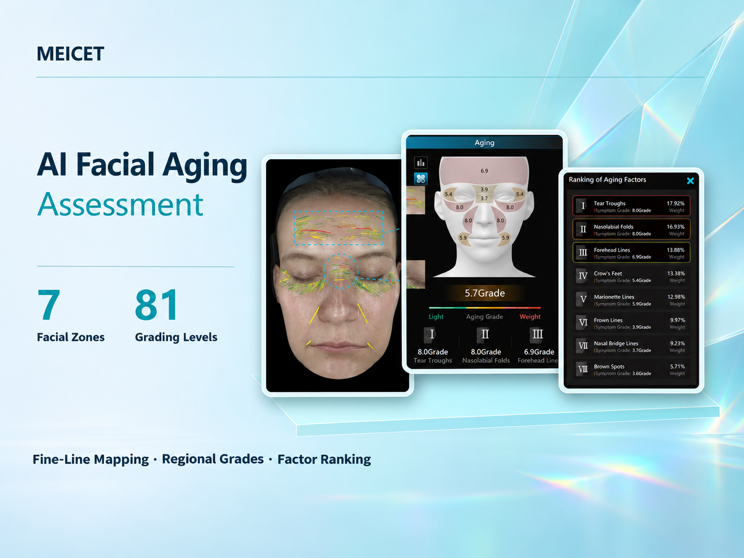
Modern professional systems employ convolutional neural networks and related deep learning architectures. The MEICET Pro-D’s AI skin aging analysis maps fine lines across seven facial zones, applies 81 grading levels, and ranks visible aging factors. The system also provides nine AI skin analysis views with illustrative aging and pigmentation simulations.
Two aspects of AI performance deserve scrutiny: training data diversity and the distinction between diagnostic output and decision support. Buyers serving multi-ethnic populations should ask vendors about the demographic composition of their training datasets and whether performance has been validated across Fitzpatrick skin types.
Matching Imaging Technology to Clinical Need
| Practice Focus | Priority Imaging Technologies | Why Less Critical |
|---|---|---|
| Skin health & pigmentation | Multi-spectral (UV, CPL, PPL) | 3D depth |
| Acne & oiliness management | Blue light, UV fluorescence, PPL | Structured-light 3D |
| Anti-aging & texture | PPL, CPL, 3D texture mapping | UV (secondary) |
| Injectable & contouring | Structured-light 3D, stereo 3D | UV (unless pigment is a concern) |
| Full-service medical aesthetics | Multi-spectral + true 3D | None—both are relevant |
Frequently Asked Questions
What is the difference between multi-spectral imaging and hyperspectral imaging in face analysis?
Multi-spectral imaging captures a small number of discrete wavelength bands (typically 4–9). Hyperspectral imaging captures dozens to hundreds of contiguous narrow bands. Hyperspectral systems are typically slower, more expensive, and rarely necessary for routine aesthetic consultations.
Do I need UV imaging in a professional face analysis machine?
UV imaging is highly valuable if your practice treats pigmentation disorders, sun damage, or acne. UV fluorescence reveals porphyrin activity and shows melasma patterns invisible under standard light.
How can I verify that a “3D” face analysis machine is actually capturing depth?
Ask the vendor to specify the depth acquisition method: structured light, stereo vision, or time-of-flight. Request a repeatability demonstration with sub-millimeter metrics.
What resolution is needed for professional skin imaging?
Pore and fine-line analysis benefits from 12MP or above. Pigment and vascular assessment depend more on spectral fidelity and polarization control than on raw pixel count.
Does ISO 13485 certification matter for a face analysis machine used in a salon?
While not legally required for cosmetic consultation tools in most jurisdictions, it indicates that the manufacturer follows rigorous design and production standards.
Can AI skin analysis replace a dermatologist’s assessment?
No. These machines are designed for cosmetic consultation support and longitudinal tracking. They do not diagnose medical conditions and should function as decision support.
What should I look for in a demo to evaluate imaging quality?
Bring a subject with known skin characteristics and compare output against your visual assessment. Ask for raw images and evaluate whether spectral views reveal structures invisible under standard lighting.
Post time: Nov-19-2026


