


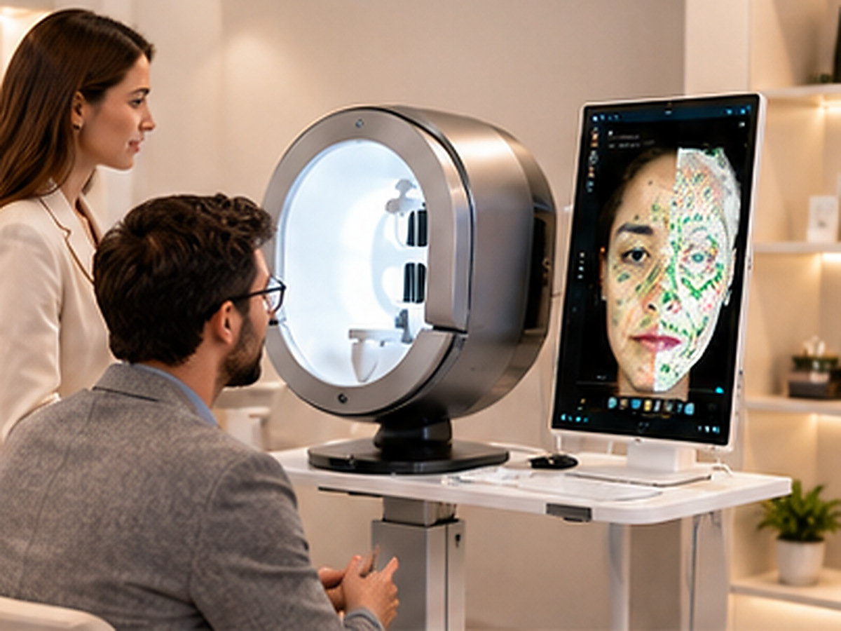
The imaging mode you need depends on which skin layer and condition you intend to assess. RGB (visible light) captures surface color and texture for baseline documentation. Cross-polarized light filters surface glare to reveal subsurface vascularity and redness. Parallel-polarized light preserves surface reflection to highlight pores, fine lines, and sebum distribution. UV light excites porphyrins produced by C. acnes and reveals deep pigmentation. The ISEMECO 3D D9 integrates all four modes into a single capture system, generating 11 spectral images from a 42-megapixel full-face sensor.
No single mode is “best.” Each spectral mode interrogates different tissue depths and structures.
Cross-polarized light is the reference standard for vascular assessment and inflammation evaluation.
Reveals porphyrins from acne bacteria and melanin absorption patterns invisible to visible light.
Parallel-polarized light serves surface texture, sebum mapping, and pore visualization.
A skin analyzer’s diagnostic value is not determined by camera resolution alone. The illumination mode determines what the camera can see. Under flat white light, a patient’s facial redness, subsurface pigmentation, and active acne inflammation blur together into a single visual impression. By switching the light source, the system isolates specific chromophores and tissue structures, converting an ambiguous image into measurable data.
Clinical literature identifies three essential light sources in facial skin imaging systems: white light, UV, and polarized light. The D9 implements this foundation while adding a distinction between parallel and cross-polarized configurations—yielding four modes that serve distinct clinical purposes.
RGB mode captures the skin as the human eye sees it—color, texture, and surface features under simulated daylight. This is the reference image against which other spectral modes are compared.
- What RGB reveals: Skin tone, surface texture, visible pores, fine lines, and superficial pigmentation.
- What RGB cannot do: Penetrate below the epidermis. Hemoglobin, basal melanin, and porphyrins remain invisible.
- Clinical role: Baseline documentation and patient communication.
Cross-polarized light filters out surface glare to reveal light that has penetrated the dermis and scattered back. This configuration is essential for assessing erythema, inflammation, and vascular conditions.
- Principle: Blocks specular reflection while transmitting diffuse reflected light from deeper layers.
- What it reveals: Facial redness, rosacea patterns, inflammatory acne lesions, and post-inflammatory erythema.
- Why it matters: Essential for laser and energy-based treatment planning.
Parallel-polarized light preserves surface reflection rather than eliminating it. This configuration enhances specular highlights, making it useful for assessing surface topography and oiliness.
- Principle: Aligned polarizers pass specular reflection from the skin surface.
- What it reveals: Pore size and density, fine lines, wrinkles, surface roughness, and sebum distribution.
- When to use it: Aesthetic consultations for texture refinement and oil control.
UV light excites fluorescence in specific skin components and reveals pigment deposition at depths invisible to visible light. This is the most clinically distinctive mode.
- Principle: UVA (320–400 nm) causes porphyrins to fluoresce orange-red and melanin to appear dark.
- What it reveals: Acne bacterial activity, deep sun damage, melasma depth, and depigmentation disorders.
- Clinical value: Quantifiable marker of C. acnes colonization for acne patients.
| Imaging Mode | Tissue Depth | Primary Structures | Best For | Limitations |
|---|---|---|---|---|
| RGB | Superficial | Color, texture | Baseline documentation | No subsurface info |
| Cross-polarized | Dermis | Hemoglobin, vascular | Redness, inflammation | No porphyrin info |
| Parallel-polarized | Surface | Pores, fine lines | Texture, oiliness | No depth info |
| UV | Epidermis to Dermis | Porphyrins, melanin | Acne activity, sun damage | Not for vascular |
The ISEMECO 3D D9 is built around a 42-megapixel binocular grating structured light camera that captures full-face images under all four spectral conditions in a single session. The system generates 11 spectral images which are processed by AI algorithms to grade features across more than 30 detection metrics.
The 3D layer adds a dimension that none of the spectral modes provide independently. The structured light system builds a topographic model with 0.2 mm accuracy, enabling volume measurement at 0.1 mL resolution. Wrinkles are measured depressions in the 3D model, with spectral data providing context about pigmentation and vascularity at that location.

For a professional practice, all four modes serve distinct clinical purposes that cannot be substituted. A system with only RGB or only UV will miss conditions that the other modes would reveal.
UV mode is uniquely valuable for visualizing porphyrin fluorescence, while cross-polarized mode is essential for assessing inflammatory erythema. They answer different questions: bacterial load vs. inflammation.
No. Cross-polarized light reveals subsurface pigmentation by removing glare, but it does not distinguish melanin depth the way UV absorption does.
It isolates the surface reflection component, producing a much clearer image of texture, pores, and sebum distribution for patient education.
The UV used is UVA at low energy, sufficient to excite fluorescence but not to cause acute photodamage. Standard precautions should still be followed.
The system switches light sources and polarizers under software control in a single session, reducing operator variability and session time.
The mode should match the treatment target. Cross-polarized for vascular, UV for pigmentation, and parallel-polarized for texture. The D9 allows comparison of the same mode across visits.
Post time: Nov-09-2026


