


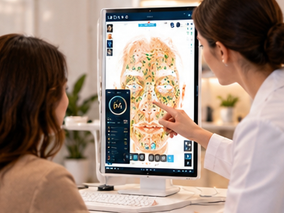
For professional skin analysis in a salon or clinic, 24 megapixels is the practical minimum, while 36–48 megapixels represents the current professional standard for capturing fine texture, small pigment spots, and pore detail without digital artifacts. A Chinese public procurement specification for facial analyzers sets the full-face pixel requirement at ≥24 megapixels, with an equivalent resolution of ≥32 megapixels as a threshold. The ISEMECO 3D D9 uses a 42-megapixel full-face sensor, while its MAX variant claims 48 megapixels. Beyond roughly 48MP, resolution gains yield diminishing clinical returns unless paired with proportionally better optics, illumination uniformity, and sensor size.
A skin analyzer’s camera resolution determines the smallest feature it can distinguish. A wrinkle that is 0.1 mm wide cannot be reliably detected if each pixel represents 0.2 mm of facial surface. The procurement specification requiring ≥24MP for full-face imaging reflects this: at typical full-face capture distances, 24MP provides sufficient pixel density to resolve fine texture and small lesions.
Beyond approximately 48MP for a full-face capture, additional megapixels produce diminishing returns for most clinical and aesthetic applications. The limiting factors become:
- Optical resolution: If the lens cannot resolve finer detail, more pixels only record blur more precisely.
- Diffraction: At small apertures, light physics limits achievable sharpness regardless of sensor resolution.
- File size and processing: A 48MP image contains roughly 2× the data of a 24MP image, increasing storage and AI inference time.
The D9’s 42MP specification sits at the upper end of the professional range—sufficient for detailed analysis without the processing overhead of 100MP+ systems.
A Chinese public procurement tender for facial analysis equipment specifies:
| Parameter | Requirement |
|---|---|
| Full-face pixels (single lens) | ≥24 megapixels |
| Image resolution | ≥8000 × 4000 or equivalent ≥32MP |
| Imaging resolution | ≥5 lp/mm |
| Illumination uniformity | ≥90% |
| Color rendering index | ≥80 |
The procurement standard requiring ≥90% illumination uniformity addresses a critical but often overlooked factor. If the center of the face is brighter than the periphery, the AI algorithm may misinterpret a lighting gradient as a skin tone variation.
A skin analyzer captures images across multiple spectral modes—RGB, cross-polarized, parallel-polarized, and UV. The D9’s specification lists solid-state LED illumination across four spectral modes, a configuration that supports consistency better than systems that rely on user-replaceable bulbs.
A 42MP image from a 1-inch CMOS sensor (D9 specification) has a different pixel pitch than a 42MP image from a smaller sensor. Larger pixels capture more light per unit area, improving signal-to-noise ratio and low-light performance.
| Specification | D9 Value | Practical Meaning |
|---|---|---|
| Full-face pixels | 42MP | At the high end of professional range; sufficient for pore and fine line analysis |
| Spectral modes | 4 (RGB, CPL, PPL, UV) | Separates surface, vascular, and deep pigment signals |
| 3D modeling accuracy | 0.2 mm | Enables contour and volume measurement |
| Image count | 11 | Multi-spectral output from a single session |
| Sensor | 1-inch CMOS | Larger than typical compact camera sensors |
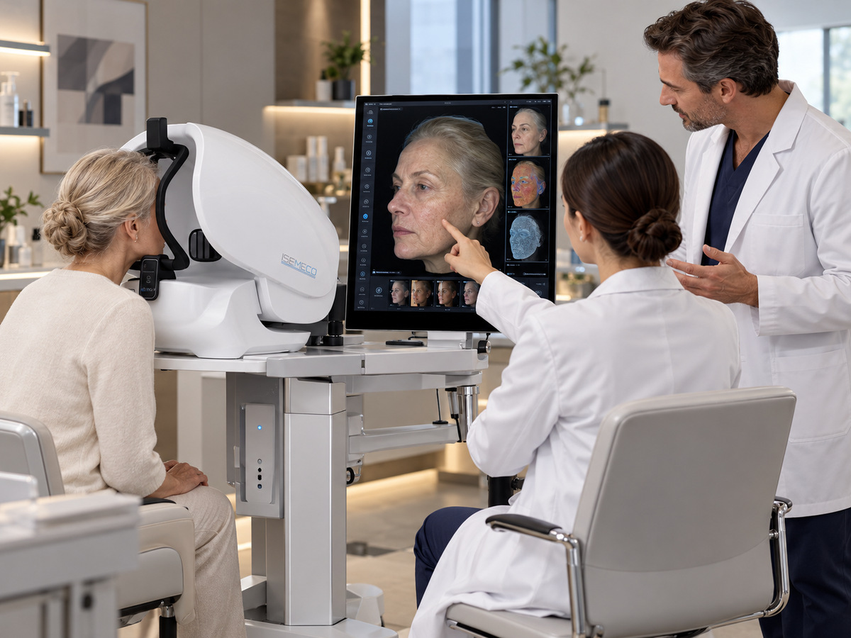
Yes, for most salon and clinic applications. The procurement threshold of ≥24MP for full-face imaging reflects the resolution needed to resolve fine texture and small pigment spots at typical capture distances.
The D9’s 42MP specification provides headroom for cropping, zooming into regions of interest, and maintaining detail across its multiple spectral modes. Higher resolution also supports the 3D reconstruction process.
Not necessarily. Wrinkle and pore detection depend on contrast and illumination angle as much as resolution. Parallel-polarized imaging may reveal pores more effectively than increasing pixel count.
The 0.2 mm figure appears consistently for 3D modeling accuracy. The 0.02 mm figure appears in one promotional source without clear definition. Buyers should ask the supplier to clarify what the 0.02 mm refers to.
RGB alone documents surface color but cannot reveal porphyrins or deep pigmentation depth. UV imaging provides information that no other spectral mode can capture, which is essential for clinical value.
A 42MP image contains roughly 1.75× the data of a 24MP image. For a system capturing 11 spectral images per session, storage and processing requirements multiply. Confirm that the system’s AI processing speed matches your throughput needs.
Request: (1) a live capture at 100% zoom to assess resolution; (2) repeat scans to evaluate consistency; (3) a sample report showing how resolution translates to metrics like wrinkle grade and pore density.
Post time: Nov-09-2026


