


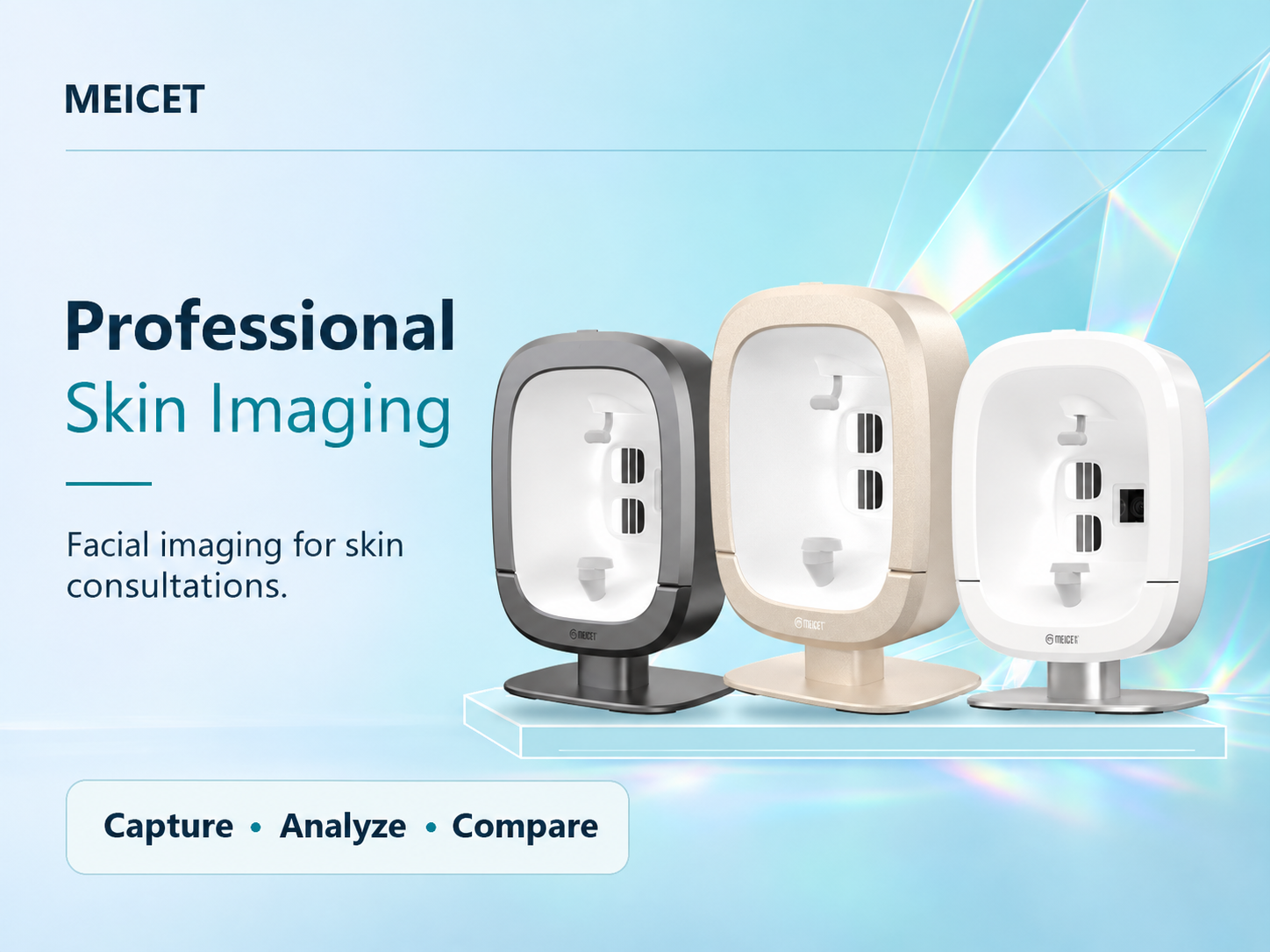
An AI face analysis machine improves facial skin assessment by replacing subjective visual judgment with objective, multi-dimensional data. The system captures images across multiple light spectra (RGB, UV, cross-polarized, parallel-polarized), reconstructs a precise 3D surface model, and applies trained algorithms to detect, classify, and grade conditions such as wrinkles, pigmentation, pores, and vascular redness. For aesthetic clinics, the core improvement is not merely sharper images—it is the conversion of a consultation from “I see some lines here” into a quantified, trackable, and visually communicable record that supports consistent treatment planning and patient education.
Key Takeaways
Objectivity Over Subjectivity
AI eliminates observer variability by applying identical detection and grading criteria to every patient, every visit, and every practitioner.
Multi-Dimensional Detection
Professional systems assess conditions across four spectral modes and generate up to 16 analysis views, revealing subsurface pigmentation and inflammation.
3D Quantification
High-end systems achieve 0.02 mm 3D resolution, enabling wrinkle depth and volume measurements that 2D photography cannot provide.
Workflow Acceleration
AI report generation in approximately 30 seconds integrates assessment into the consultation without creating bottlenecks.
A 2025 study on AI skin feature detection reported a mean average precision (mAP@0.5) of 0.833 for wrinkle and pore detection under optimized conditions, indicating that algorithmic identification of subtle textural features is technically robust. The practical question for clinic buyers is whether this technical capability translates into better consultations, more consistent treatment selection, and measurable patient outcomes.
What Exactly Does “AI” Add to a Face Analysis Machine?
AI adds three capabilities that conventional imaging cannot deliver: automated feature detection, standardized severity grading, and predictive simulation. Without AI, a face analysis machine produces images that a practitioner must interpret manually. With AI, the system performs the first-pass analysis, identifies relevant features, ranks concerns by severity, and presents structured findings.
Automated Feature Detection and Grading
AI models trained on annotated skin image datasets detect features such as wrinkles, pores, and pigmentation with increasing accuracy. The 2025 study cited above used a YOLOv8s detector optimized with particle swarm optimization to achieve mAP@0.5 of 0.833 on a high-quality test subset. While laboratory performance does not guarantee clinical accuracy across all skin types and lighting conditions, the result demonstrates that AI can reliably localize subtle features that human observers might overlook or grade inconsistently.
The practical benefit for a clinic is consistency. A practitioner assessing ten patients in a day may unconsciously shift criteria between the first and the last consultation. An AI system applies the same detection thresholds and grading scales to every image, creating a comparable record across visits.
Predictive Aging Simulation
AI-assisted aging simulation projects how current skin patterns may evolve over 5–10 years, using parameters such as wrinkle progression, volume loss, and pigmentation accumulation. MEICET’s clinical materials describe the D9 platform’s aging forecast as covering ages 18 to 80, with 8 anti-aging dimensions and 81 aging grades. These projections are illustrative tools for patient education, not deterministic predictions.
AI in aesthetic face analysis detects and quantifies visible features; it does not diagnose disease. Research on AI skin disease algorithms has reported skin cancer sensitivity of 78.2% and specificity of 88.0% in validation studies. When a lesion appears suspicious, the machine’s output is irrelevant; the patient requires dermatological evaluation.
How Does Multi-Spectral Imaging Improve Detection Accuracy?
Multi-spectral imaging improves detection accuracy by capturing skin information at different depths and contrast mechanisms, revealing conditions that a single visible-light image cannot distinguish.
| Spectral Mode | What It Captures | Clinical Relevance |
|---|---|---|
| RGB (Visible Light) | Surface color, texture, visible pores | Baseline documentation, patient communication |
| Cross-polarized | Subsurface redness, dermal pigment, vascularity | Rosacea assessment, melasma depth evaluation |
| Parallel-polarized | Surface shine, texture irregularities | Sebum assessment, skin smoothness grading |
| UV Light | Porphyrins, sun damage, melanin distribution | Photodamage detection, acne bacteria visualization |
How Does 3D Modeling Change Assessment Compared to 2D?
3D modeling changes assessment by converting visual impressions of depth into measurable quantities. A 2D photograph shows that a nasolabial fold exists; a 3D model measures whether the fold is 0.3 mm or 1.2 mm deep.
| Assessment Feature | 2D Imaging | 3D Modeling | Clinical Impact |
|---|---|---|---|
| Wrinkle presence | Visible | Measurable depth (mm) | Distinguishes superficial vs. deep folds |
| Facial volume | Shadow patterns only | Quantified volume difference (0.1 mL) | Validates filler outcomes |
| Contour / Asymmetry | Perspective-dependent | Measurable angles and proportions | Supports structural treatment planning |
AI-Powered Assessment Workflow
How Should Clinics Evaluate AI Claims?
Questions to Ask During Evaluation:
- Can you demonstrate detection on a patient with melasma or rosacea?
- What is the 3D resolution specification? (e.g., D9MAX’s 0.02 mm)
- How does the system handle Fitzpatrick IV–VI skin?
- Does the report link findings to treatment categories?
Red Flags in AI Marketing:
- Claims of diagnostic capability for skin disease without regulatory clearance.
- Vague accuracy claims without dataset validation.
- No demonstration on real patients with common aesthetic concerns.
Frequently Asked Questions
What does an AI face analysis machine actually measure?
It measures visible and subsurface skin features: wrinkle depth, pigmentation distribution, pore size, vascular redness, skin texture, and facial volume. It does not measure hydration or elasticity unless equipped with a separate probe.
Can an AI face analysis machine tell me if a spot is cancerous?
No. These are not diagnostic devices for skin cancer. Any suspicious lesion requires evaluation by a qualified dermatologist.
How long does an AI skin analysis take?
Current professional systems generate complete AI reports in approximately 30 seconds, with image capture taking only seconds.
Do I need special lighting for the machine to work accurately?
No external lighting control is required. Professional machines use internal solid-state LED arrays under controlled spectral modes.
Is AI face analysis worth the investment for a small clinic?
The value depends on consultation volume and treatment mix. Clinics performing injectables or energy-based treatments benefit most from objective documentation and tracking.
Post time: Nov-17-2026


