


A 3D skin analyzer works by projecting a structured light pattern onto the face and capturing its deformation across two camera viewpoints to reconstruct facial topography with sub-millimeter precision. The ISEMECO D9 uses binocular grating structured light to achieve 0.2mm modeling accuracy and 42 million full-face pixels, enabling volume measurement to 0.1ml precision and an 8-dimension aging analysis covering forehead lines through brown spots. The technology stack combines four spectral imaging modes (RGB, cross-polarized, UV, parallel-polarized) with AI deep learning to convert geometric and spectral data into quantified clinical outputs. Buyers should understand three technical layers: how the 3D reconstruction actually works, what the spectral modes reveal that RGB cannot, and where the AI adds value versus where it simply automates pattern recognition.
Key Takeaways
Structured light is the core 3D mechanism: a known grating pattern is projected onto the face; depth is calculated from how the pattern bends across the surface.
The D9’s 0.2mm accuracy enables clinical-grade volume tracking, including 0.1ml precision for filler and contour assessment.
Spectral modes reveal subsurface information: cross-polarized shows vascular patterns; UV highlights porphyrins and pigmentation depth.
AI classifies reconstructed data: the aging model is trained on big data to grade 8 dimensions and predict trajectories.
Fitzpatrick I–VI calibration adjusts classification thresholds for different melanin levels, addressing dermatology AI failure modes.
The 3D Reconstruction Mechanism: How Structured Light Measures Facial Topography
A 3D skin analyzer does not “take a picture” in the conventional sense. It projects a known pattern onto the face and measures how that pattern deforms when it strikes three-dimensional surfaces.
The ISEMECO D9 uses binocular grating structured light—a technique that projects a grating pattern from one angle and captures it from two camera viewpoints. When the grating lines hit a curved surface (a cheek, a nose bridge, a wrinkle), they bend. The amount of bending, measured against the known projection geometry, encodes depth. The two-camera configuration resolves ambiguities that a single-camera setup cannot.
The result is a point cloud or mesh representing facial topography. The D9 reports this reconstruction with 0.2mm modeling accuracy. For context, 0.2mm is approximately the thickness of two sheets of paper—fine enough to detect subtle changes in wrinkle depth or contour that would be invisible in a flat photograph.
This geometric model is what enables the D9’s volume difference calculation to report changes with 0.1ml precision. A 2D image cannot measure volume because it contains no depth data; the structured-light reconstruction is the prerequisite for any volumetric claim.
The Spectral Layer: What Four Imaging Modes Reveal
The D9 captures images under four spectral conditions: RGB (visible light), cross-polarized, parallel-polarized, and UV. Each mode interrogates different tissue depths and structures.
- •RGB (visible light) captures surface color and texture as the eye sees them—useful for baseline documentation but limited in depth penetration.
- •Cross-polarized light filters out surface glare and selectively captures light that has penetrated into the dermis and scattered back. This mode reveals subsurface vascular patterns and redness that surface photography obscures. Clinical skin imaging research confirms that cross-polarized configurations are essential for assessing erythema and vascular conditions.
- •Parallel-polarized light preserves surface reflections, making it useful for assessing sebum distribution and surface shine.
- •UV light excites porphyrins produced by C. acnes bacteria and highlights pigmentation depth—sun damage that sits below the surface appears differently under UV than under visible light.
The D9’s software integrates these modes with the 3D topography. A wrinkle is not just a dark line in a photo; it is a measured depression in the 3D model, assessed alongside the spectral signature of the surrounding tissue.
The AI Layer: What the Algorithm Does—and Does Not Do
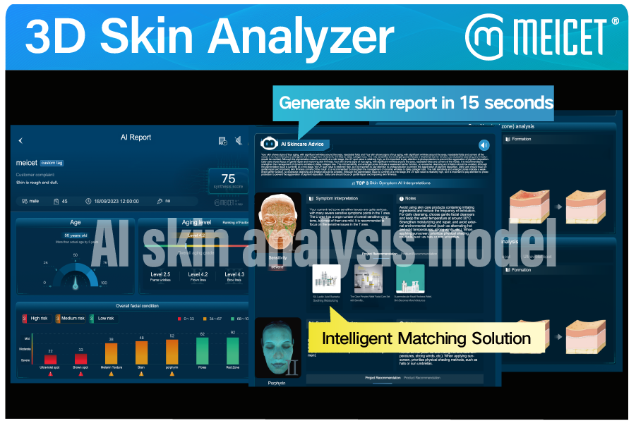
The AI in a 3D skin analyzer operates on reconstructed data, not on raw pixels. The D9’s aging model is described as using AI deep learning technology built on big data skin images to construct an 8-dimension aging index covering forehead lines, glabellar lines, crow’s feet, periorbital lines, laugh lines, marionette lines, corner of mouth lines, and brown spots.
What this means in practice: the algorithm takes the geometric and spectral data and assigns grades (mild, moderate, severe) to each dimension. It can also perform aging weight ranking and trajectory prediction—projecting how a patient’s aging profile may evolve.
What the algorithm does not do is diagnose disease. The D9 is positioned for aesthetic and dermatological assessment, not for skin cancer detection. Buyers should not conflate “AI skin analysis” with “diagnostic AI.” The FDA regulates AI-enabled medical devices based on intended use and risk; a device claiming diagnostic capability enters a different regulatory pathway than one claiming aesthetic assessment.
The Fitzpatrick I–VI calibration addresses a well-documented weakness in dermatology AI: models trained predominantly on lighter skin tones can misclassify conditions on darker skin. The D9’s explicit skin-type adjustment is a software-level mitigation, not a hardware fix. Buyers serving diverse populations should verify whether the calibration is automatic (algorithm detects skin type) or manual (operator selects), as manual selection introduces user-dependence.
Technical Specifications Buyers Should Understand
| Specification | D9 Reported Value | What It Means for the Buyer |
|---|---|---|
| 3D structured light | Binocular grating | Depth from pattern deformation, not stereo estimation |
| Modeling accuracy | 0.2mm | Detection of subtle contour and wrinkle-depth changes |
| Volume precision | 0.1ml | Quantified filler and tissue-change tracking |
| Full-face pixels | 42 million | High-resolution surface texture capture |
| Spectral modes | RGB, cross, parallel, UV | Multi-depth tissue assessment |
| CMOS sensor | 1 inch | Larger sensor area for light capture and reduced noise |
| Interface | USB 3.0 DC | Data transfer speed for high-resolution imaging |
| Instrument weight | 19.5 kg | Workstation-class device, not portable |
Source: MEICET product page for ISEMECO D9
The 1-inch CMOS sensor matters for image quality: a larger sensor captures more light per pixel, reducing noise in dim spectral modes (especially UV). The USB 3.0 interface is necessary for transferring 42-megapixel images without unacceptable delay.
The 19.5 kg weight signals that this is a fixed-installation device. Buyers planning mobile or multi-room use should factor in the logistics of moving and recalibrating a workstation-class instrument.
Market Context: Where the D9 Sits
MEICET has positioned the D9 for dermatologists, cosmetic surgeons, and premium skin care centers, presenting it at international exhibitions including Cosmoprof Bologna, AMWC Monaco, and AMWC Dubai. The company reports 5,000+ clinics served globally and CE/FDA certifications.
A MEICET press release describes the D9’s AI diagnostic engine as including predictive aging modeling that projects skin conditions 5–10 years ahead using 12 clinical parameters, and a treatment simulator that visualizes potential procedure outcomes in real time. These are marketing claims that buyers should verify through product demonstration and reference customer contact, not accept at face value.
The competitive landscape includes other 3D skin imaging systems (Quantificare’s LifeViz line, Canfield’s VECTRA, and Miravex’s Antera) that use different reconstruction methods. Buyers comparing systems should ask each supplier to demonstrate repeatability—the same face scanned twice should produce consistent measurements—rather than relying on single-scan demonstrations that cannot reveal calibration drift.
FAQ
It projects a structured light pattern (the D9 uses binocular grating) onto the face and captures the pattern from two viewpoints. The deformation of the pattern encodes surface depth. This is fundamentally different from stereo photography or time-of-flight sensors, which have different accuracy profiles and failure modes.
Modeling accuracy describes how faithfully the 3D reconstruction captures surface geometry—a 0.2mm error means the reconstructed surface is within 0.2mm of the true surface. Volume precision describes the smallest volume change the software can reliably report—0.1ml. They are related but distinct specifications.
The D9 captures four spectral modes (RGB, cross-polarized, parallel-polarized, UV). The product page does not specify whether capture is simultaneous or sequential. Buyers should ask during demonstration: sequential capture with patient movement between modes can introduce alignment errors.
The D9 includes Fitzpatrick I–VI adjustment to “avoid errors” across skin types. The critical question is whether this adjustment is automatic or manual. Automatic detection reduces operator error and ensures consistent application.
The eight dimensions are forehead lines, glabellar lines, crow’s feet, periorbital lines, laugh lines, marionette lines, corner of mouth lines, and brown spots. Each is graded based on the 3D geometry and spectral data, providing treatment targets rather than just a single score.
The D9’s published materials describe aesthetic and aging analysis, not diagnostic claims. Buyers should confirm the device’s intended use statement matches their clinical workflow and that regulatory clearances (CE, FDA) cover the claims they plan to make.
Three things: repeatability (scan the same person twice), Fitzpatrick calibration (test on a darker-skinned volunteer), and report output (does the generated report contain the specific numbers and recommendations your clinic needs?).
Post time: Oct-23-2026


