


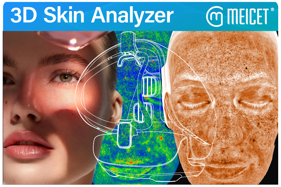
For professional skin assessment, 3D skin analyzers are generally superior to 2D systems because they provide depth information that 2D photography fundamentally cannot capture. A 3D analyzer like the ISEMECO D9 uses binocular grating structured light to reconstruct facial topography with 0.2mm modeling accuracy, enabling quantitative measurement of volume changes to 0.1ml precision—a capability that 2D imaging lacks entirely. Clinical research in facial anthropometry confirms that 3D methods achieve acceptable accuracy while 2D photography shows discrepancies exceeding clinically acceptable limits. However, 2D analyzers remain viable for basic surface assessment and budget-constrained settings.
The Fundamental Difference: What 3D Captures That 2D Cannot
A 2D skin analyzer captures a flat image—essentially a high-resolution photograph with software analysis layered on top. The image contains color information (redness, pigmentation, surface shine) and texture information (pores, fine lines visible at the surface). What it does not contain is depth: the physical topography of the face, the volume of the cheeks, the projection of the chin, or the actual three-dimensional shape of a wrinkle.
A 3D skin analyzer reconstructs this missing dimension. The ISEMECO D9 uses binocular grating structured light—a technique that projects a known pattern onto the face and calculates depth from the pattern’s deformation across two camera viewpoints. The result is a digital model of facial topography that can be measured, compared, and quantified in physical units.
Clinical Evidence: 3D Accuracy Versus 2D Limitations
The accuracy advantage of 3D imaging is documented in clinical research, particularly in facial anthropometry—the measurement of facial dimensions and proportions. A 2023 clinical study published in the Journal of Clinical Medicine compared a dual-structured light 3D facial scanner against 2D photography. The study concluded that the 3D method reported acceptable accuracy values, while the 2D method showed discrepancies over the clinically acceptable limits.
- Volume changes after filler or energy-based treatments
- Facial symmetry in three dimensions
- Wrinkle depth (2D shows darkness, not physical depth)
- Contour irregularities that depend on viewing angle
What the ISEMECO D9 Provides: 3D Capabilities in Specification
| Specification | Reported Value | What It Enables |
|---|---|---|
| 3D structured light | Binocular grating | Depth reconstruction from pattern deformation |
| Modeling accuracy | 0.2mm | Detection of subtle topographic changes |
| Volume calculation | 0.1ml precision | Quantification of filler and tissue changes |
| Full-face pixel count | 42 million | High-resolution surface texture capture |
When 2D Remains the Appropriate Choice
Despite the clinical advantages of 3D, 2D analyzers remain appropriate in specific scenarios: Budget-constrained settings, basic surface assessment (redness/pores), high-volume screening without treatment planning, and portable or space-constrained environments. The decision framework is: “3D is better when the clinical or commercial workflow requires depth information.”
| Capability | 2D Analyzer | 3D Analyzer (D9 Class) | Clinical Relevance |
| Wrinkle assessment | Visual/color-based | Depth-measured | 3D enables quantification |
| Volume change | Not possible | 0.1ml precision | Essential for filler tracking |
| Facial symmetry | 2D only | 3D contour analysis | 3D detects depth asymmetry |
Post time: Oct-23-2026


